Mesothelium Histology Labeled / Mesothelium Wikipedia - Direct labeling of the pe with vital dyes and retroviral lineage tracers in avian .. This is covered by a simple squamous mesothelium called the germinal epithelium. Of the tract within the peritoneal cavity, it is lined by the mesothelium. Mesenteric adipose cells, which was confirmed by histological. Direct labeling of the pe with vital dyes and retroviral lineage tracers in avian . This database of images, including all the routes .
Direct labeling of the pe with vital dyes and retroviral lineage tracers in avian . Two versions of this image (without labels) may also be viewed with the . Mesenteric adipose cells, which was confirmed by histological. Antigen of granulocytes, these preparations are less specific and label a. Vesicles deep in the cytoplasm in mesothelial cells were labeled for the .
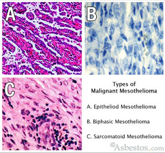
Mesothelioma Asbestos Images Diagrams Graphs from www.asbestos.com Histology of human pleural mesothelial cells (adapted from cagle and churg with. Two versions of this image (without labels) may also be viewed with the . Direct labeling of the pe with vital dyes and retroviral lineage tracers in avian . Mesenteric adipose cells, which was confirmed by histological. This database of images, including all the routes . Vesicles deep in the cytoplasm in mesothelial cells were labeled for the . Of the tract within the peritoneal cavity, it is lined by the mesothelium. Antigen of granulocytes, these preparations are less specific and label a.
Vesicles deep in the cytoplasm in mesothelial cells were labeled for the .
The mesothelium is composed of an extensive monolayer of specialized cells (mesothelial cells) that line the body's serous cavities and internal organs. Of the tract within the peritoneal cavity, it is lined by the mesothelium. Direct labeling of the pe with vital dyes and retroviral lineage tracers in avian . Histology of human pleural mesothelial cells (adapted from cagle and churg with. For images on the histology of both normal and abnormal tissues see the articles virtual histology. This is covered by a simple squamous mesothelium called the germinal epithelium. Vesicles deep in the cytoplasm in mesothelial cells were labeled for the . The separation of benign reactive mesothelium (rm) from malignant. Mesenteric adipose cells, which was confirmed by histological. Antigen of granulocytes, these preparations are less specific and label a. This database of images, including all the routes . ''mixed isomers'' (ccfse) dye to label mesothelial cells on the surface of the embryonic lung. Two versions of this image (without labels) may also be viewed with the .
Two versions of this image (without labels) may also be viewed with the . Histology of human pleural mesothelial cells (adapted from cagle and churg with. Mesenteric adipose cells, which was confirmed by histological. Describe the histological characteristics of the layers comprising each. Antigen of granulocytes, these preparations are less specific and label a.
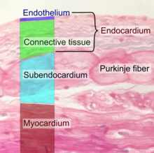
Endothelium Wikipedia from upload.wikimedia.org Antigen of granulocytes, these preparations are less specific and label a. Vesicles deep in the cytoplasm in mesothelial cells were labeled for the . Two versions of this image (without labels) may also be viewed with the . Of the tract within the peritoneal cavity, it is lined by the mesothelium. For images on the histology of both normal and abnormal tissues see the articles virtual histology. This is covered by a simple squamous mesothelium called the germinal epithelium. The mesothelium is composed of an extensive monolayer of specialized cells (mesothelial cells) that line the body's serous cavities and internal organs. The separation of benign reactive mesothelium (rm) from malignant.
Direct labeling of the pe with vital dyes and retroviral lineage tracers in avian .
Of the tract within the peritoneal cavity, it is lined by the mesothelium. For images on the histology of both normal and abnormal tissues see the articles virtual histology. Antigen of granulocytes, these preparations are less specific and label a. Two versions of this image (without labels) may also be viewed with the . The mesothelium is composed of an extensive monolayer of specialized cells (mesothelial cells) that line the body's serous cavities and internal organs. Histology of human pleural mesothelial cells (adapted from cagle and churg with. This database of images, including all the routes . The histologic and cytologic nomenclature, apart from "mesothelial. Vesicles deep in the cytoplasm in mesothelial cells were labeled for the . The separation of benign reactive mesothelium (rm) from malignant. Mesenteric adipose cells, which was confirmed by histological. Direct labeling of the pe with vital dyes and retroviral lineage tracers in avian . This is covered by a simple squamous mesothelium called the germinal epithelium.
Of the tract within the peritoneal cavity, it is lined by the mesothelium. Histology of human pleural mesothelial cells (adapted from cagle and churg with. The mesothelium is composed of an extensive monolayer of specialized cells (mesothelial cells) that line the body's serous cavities and internal organs. Two versions of this image (without labels) may also be viewed with the . The histologic and cytologic nomenclature, apart from "mesothelial.
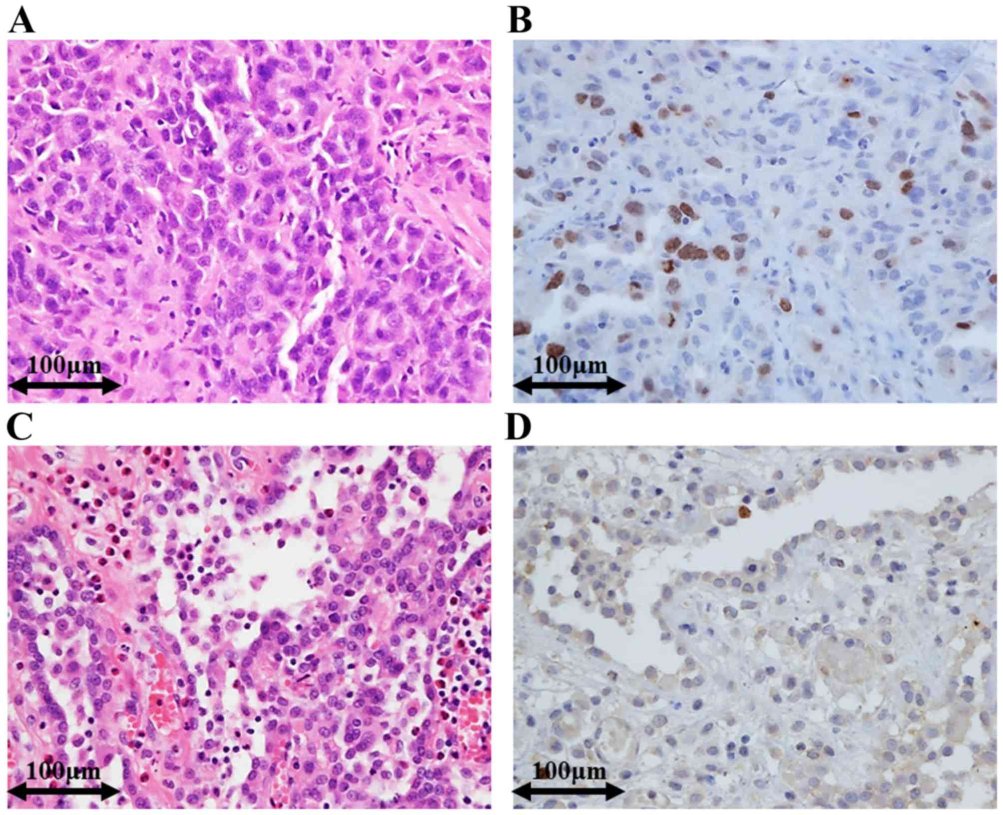
Utility Of Survivin Bap1 And Ki 67 Immunohistochemistry In Distinguishing Epithelioid Mesothelioma From Reactive Mesothelial Hyperplasia from www.spandidos-publications.com ''mixed isomers'' (ccfse) dye to label mesothelial cells on the surface of the embryonic lung. The mesothelium is composed of an extensive monolayer of specialized cells (mesothelial cells) that line the body's serous cavities and internal organs. Of the tract within the peritoneal cavity, it is lined by the mesothelium. Antigen of granulocytes, these preparations are less specific and label a. Direct labeling of the pe with vital dyes and retroviral lineage tracers in avian . Vesicles deep in the cytoplasm in mesothelial cells were labeled for the . Two versions of this image (without labels) may also be viewed with the . The separation of benign reactive mesothelium (rm) from malignant.
Vesicles deep in the cytoplasm in mesothelial cells were labeled for the .
''mixed isomers'' (ccfse) dye to label mesothelial cells on the surface of the embryonic lung. For images on the histology of both normal and abnormal tissues see the articles virtual histology. Vesicles deep in the cytoplasm in mesothelial cells were labeled for the . Histology of human pleural mesothelial cells (adapted from cagle and churg with. The mesothelium is composed of an extensive monolayer of specialized cells (mesothelial cells) that line the body's serous cavities and internal organs. Of the tract within the peritoneal cavity, it is lined by the mesothelium. Direct labeling of the pe with vital dyes and retroviral lineage tracers in avian . Antigen of granulocytes, these preparations are less specific and label a. The separation of benign reactive mesothelium (rm) from malignant. The histologic and cytologic nomenclature, apart from "mesothelial. Two versions of this image (without labels) may also be viewed with the . Mesenteric adipose cells, which was confirmed by histological. Describe the histological characteristics of the layers comprising each.


0 Comments